ATOS – Your knee specialist in Germany

In the ATOS clinics you can rely on the expertise of our highly specialised and renowned physicians, who are among the best knee specialists in Germany.

The knee is the largest joint of the human body – and is very complicated and prone to injury. This complex part of the body is composed of the femur, the tibial head and the kneecap. The inner and outer meniscus are located between the upper and lower leg bones, while the knee joint is surrounded by a capsule. This produces the synovial fluid which is like the lubricant for joints. The stability of the knee is ensured by the anterior and posterior cruciate ligaments and the equally important inner and outer ligaments located on the inside and outside of the knee. To prevent the individual joints from rubbing against each other painfully, the cartilage forms a protective elastic layer between the bones.

There are many other causes of knee problems – and many therapeutic and surgical options

Most commonly, knee injuries are due to cruciate ligament and meniscus tears. A meniscal tear often manifests itself in acute pain, swelling, and blockage of the knee joint. A tear of the meniscus can either be due to signs of wear or trauma, for example a sports injury. Although meniscal tears can occasionally be treated conservatively at first, meniscus surgery is usually necessary. As a rule, this is done arthroscopically, i.e. minimally invasive.

The anterior or posterior cruciate ligament often ruptures due to large force, or over-rotation or extension of the knee joint. When this has occurred, treatment is absolutely necessary – on the one hand to restore full mobility, and on the other hand to prevent long-term problems such as knee osteoarthritis (gonarthrosis). There are approaches to conservative treatment of cruciate ligament damage, but in most cases, cruciate ligament surgery is recommended. Like meniscus surgery, the surgical treatment of the cruciate ligament is usually arthroscopic.

In addition, there are many other possible knee injuries, such as a tear of the inner ligament, cartilage damage and gonarthrosis as mentioned above. Implantation of an artificial knee joint (knee TEP) is usually the last solution here. Knee endoprosthesis of this kind can restore painless mobility.

Our treatments in detail

Doctors refer to osteoarthritis in the knee joint as gonarthrosis. This is a deterioration of the articular cartilage, which is the most common cause of knee joint disease. Deformity of the legs, constant improper loading, rheumatism, metabolic diseases or the consequences of an injury can damage the articular cartilage.

Definition
The development of osteoarthritis, area-wide cartilage wear, is easily caused by cartilage damage but can also result from unilaterally high stress (e.g. in tilers, high-performance athletes), congenital misalignment of the legs (e.g. O- or X-legs) or poor genetic dispositions. An unstoppable wear process begins, and the knee joint deforms and becomes inflamed.

For very pronounced X-legs there is a one-sided strain on the outside of the knee joint and for an O-leg on the inside of the knee joint. This promotes early cartilage wear. Such deformities are not always innate; they can also be caused by fractures that have grown together incorrectly.

If the gonarthrosis is advanced, and the articular cartilage is severely attacked, non-surgical treatments are often no longer sufficient. An artificial joint then offers the chance of a new life of mobility.

Symptoms
The symptoms of knee osteoarthritis are very different in patients. Initially, the joints only hurt during exercise but later also at rest. Pain at rest is an indication of advanced injury. Limitations of movement, swelling, muscle tension, feelings of instability and rubbing sounds are typical symptoms of gonarthrosis.

Diagnosis
There is no single, self-evident typical clinical symptom of osteoarthritis. Rather, there are several typical but nonspecific clinical signs that indicate the presence of osteoarthritis. These include pain, impaired function, crepitus (joint crunching), palpable osteophytes (bony outbreaks), swelling, effusion, axial deviation and instability. In most cases, an X-ray is done if the disease is suspected. And the damage is assessed. An ultrasound examination is also often performed. Muscles, ligaments and joint fluids can be accurately represented.

Conservative treatment
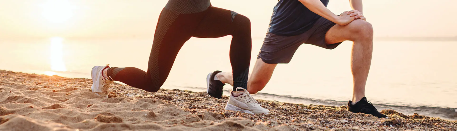
The aim of conservative therapy is to alleviate knee pain and favourably influence the further course of the disease. Osteoarthritis can often be treated conservatively (e.g. with injections) depending on the extent. This is aimed at alleviating the symptoms with movement, strengthening, physical and/or medical therapy. Cycling is considered essential. Injection therapies with hyaluronic acid, ACP or orthokin (autologous blood therapy) often lead to a reduction in pain and thus an improvement in the joint situation.

Surgical treatment

Knee arthroscopy
Arthroscopy is a minimally invasive form of knee joint exploration. The arthroscope used in this case consists of a probe with a miniature camera and two hoses, via which a rinsing liquid can be filled into the joint and sucked out. The camera transmits images from the interior of the joint to a monitor. If damage to the joint develops during the examination, it can usually be treated directly during the procedure. For this purpose, additional instruments are introduced via additional skin incisions. It is also possible to take tissue samples to have them examined in a histological examination. If no treatable changes can be detected during the exploration, the instruments are removed again, the flushed liquid is sucked out and the skin incisions are taken care of. The arthroscopy usually takes about 30 minutes, but depending on the extent of the damage to be repaired, the time may vary slightly.

Knee replacement
Knee endoprosthesis (knee TEP) is useful whenever the functionality and flexibility of the joint cannot be restored with conservative treatment and the patient suffers from persistent pain or restricted mobility.

Depending on the clinical picture and its severity, different types of artificial joints are used. An endoprosthesis is understood to mean the replacement of the individual parts of the joint by artificial materials. This operation requires intensive follow-up, so that the new joint can consolidate optimally in the bone. Physiotherapy helps to accustom the artificial joint to everyday movements. Due to the many years of experience of our ATOS knee specialists in the use of knee endoprostheses (knee TEPs), very high patient satisfaction can be expected with this operation.

In surgical treatment, one differentiates between two prosthesis types, which can be used depending on the type of osteoarthritis:

  • One-sided prosthesis (sled prosthesis/unicondylar). This type of prosthesis requires wear in the inner joint section and an otherwise largely healthy knee joint in order to achieve an optimal result.
  • Two-sided prosthesis (total endoprosthesis/TEP/bicondylar). This type of prosthesis is implanted with developed wear in several compartments of the knee joint.

In addition to the conventional surgical methods, there are also soft tissue-sparing minimally invasive techniques in which the muscles are not severed, but only pushed aside. During surgery, the diseased knee joint is replaced by an artificial joint. The implantation takes about 90 minutes and can be done in full or partial anesthesia. Partial anesthesia is less stressful for the body.

Rehabilitation – Time and methodology
The rehabilitation begins right after the operation, because early mobilisation helps the habituation and strengthening of the surrounding muscles. Walking training, stair climbing etc. are incorporated as soon as possible in the rehabilitation program. The aim of the physiotherapeutic treatment is also to correct the improper stress levels arising due to the restriction in movement over the past months and years. The physiotherapy starts right after the operation. After learning to walk under-arm crutches, the patient will climb light stairs after 4-5 days. A 3-4 week rehabilitation is needed. After 4-6 weeks you should be able to fully load the knee TEP. The artificial joints are stable for more than 20 years if treated correctly.

The kneecap, or patella, is a disc-shaped bone in front of the knee joint. It is attached on tendons, muscles and ligaments. Instabilities of the kneecap are painful and cause untreated secondary damage. Patella instabilities can be discovered early by experienced ATOS clinicians through various manipulations and special movements of the knee and treated safely without any consequential damage.

Definition
Patellar instability, kneecap dislocation and their precursors are a very common condition in daily practice, especially in adolescents. Since the kneecap has no bony guidance and slides only in a groove of the femur, it is prone to injury. With a constitution-based malformation of the patella, the plain bearing or the holding tendons, muscles and ligaments are too weak and cause patellar instability. Patellar instability also results from the tearing of the inner knee ligament (MPFL – medial patello-femoral ligament) as the result of a dislocation of the kneecap. It can also lead to a circulatory disorder and the death of bone areas, whereby the overlying cartilage can also be destroyed.

Contributing factors include:

  • Deformation of the legs (X-leg position)
  • Too flat (hypo or dysplastic) manifestation of the patellar surface and the articular surface of the thigh
  • General connective tissue weakness
  • Muscular dysbalance with a preponderance of the lateral muscle parts

Symptoms
Patellar instability patients usually complain of pain in the front knee. Pain when walking downhill is typical. As a consequence of an acute dislocation, a sudden sagging of the knee is usually described. Often, a deformation of the knee is recognisable, with the kneecap is shifted to the outside. The knee is protected in the bent position and the mobility of the knee joint is limited. Articular effusion causes pressure sensitivity. Dislocation of the kneecap can occur repeatedly.

Diagnosis
After a consultation with the patient (anamnesis), our orthopaedic surgeons can diagnose instability through various movements and special motions of the knee, which are documented by imaging techniques. It is important to distinguish harmless problems in the growth phase from real instabilities and imbalances. A targeted problem analysis which goes beyond the knee joint itself is often necessary here. X-rays or magnetic resonance imaging (MRI) are used to visualise injuries to the kneecap. If cartilage is affected, arthroscopy is often performed.

Conservative treatment
In conservative therapy, after an initial immobilisation, a specific physiotherapy is started, in which the medial muscle group is strengthened to stabilise the patella. Knee braces can be helpful at first.

Surgical treatment
If surgical treatment for the stabilisation and prevention of consequential damage to the articular cartilage becomes necessary after acute dislocation of the kneecap or chronic instability, a combination of individual measures is often necessary.

The central therapeutic approach is the reconstruction of the inside knee ligament (MPFL) as the most important passive stabiliser of the knee joint. Similar to a cruciate ligament replacement surgery, this operation achieves optimal retention of the patella in extension and flexion. In individual cases, combination with further individual stabilising measures is required.

With cartilage splits within the joint, risk of another kneecap dislocation and an athletic activity level of the patient arthroscopic supply to the cartilage or bone damage is required in addition to surgery on the inside knee tendon.

Rehabilitation  – Time and methodology
In order to avoid renewed dislocation, muscle building training is a central point of the therapy. Stronger flexion is possible after 4 to 6 weeks. Full recovery is expected after about 3 months.

The articular cartilage is a special tissue. It ensures that the components of a joint can move against each other almost without friction. It is so special that Stern magazine called it “white gold” in its October 2017 issue. Since the cartilage has neither nerves nor pain fibres, we can move painlessly. However, damaged cartilage reveals itself to us very late.

Definition
In a joint, two rigid bones are movably connected. To prevent rubbing of the bones, these contact surfaces are covered with cartilaginous tissue. It is a vascular-free supporting tissue consisting of cartilage cells (chondrocytes). These form collagen fibres, which are crosslinked together. A lot of water can be bound between the fibres to allow for elasticity and to protect against pressure loading. Due to the large amount of water that can be bound between the cartilage cells, the articular cartilage is very pressure-elastic. Lubricity and impact resistance is thereby ensured within the joint. There are no blood vessels in the cartilage, so the supply of nutrients occurs through the cartilaginous skin and synovial fluid.

Cartilage damage inhibits function and is painful. In the form of osteoarthritis, it is among the most common and significant chronic diseases of adults, especially in old age. Cartilage damage is either traumatic, for example due to a sports injury or degenerative in the context of stress, overweight and recurring micro injuries. Since the cartilage has no possibility of its own regeneration, untreated cartilage damage inevitably leads to the complete destruction of the joint, which can only be treated by the installation of an artificial joint, an endoprosthesis.

Symptoms
Cartilage is completely insensitive to pain. Illnesses or damage do not trigger alarm signals in the form of pain as occurs for example with the skin. Injuries are only noticed when the cartilage damage has occurred and pain from concomitant damage occurs. Therefore, early detection is extremely important, because corrective measures can only be initiated if there is still cartilage substance. If the cartilage layer is worn away or destroyed too far, only joint replacement measures remain. In already damaged cartilage, swelling of the knee often occurs due to the retention of fluids.

Diagnosis
Nuclear magnetic resonance imaging (MRI) is the method of choice for detecting early damage. Modern MRI diagnostics delivers high-resolution tomograms of all cartilage in the human body without radiation exposure to the patient and within a very short time. In addition, an MRI can provide information on specific cartilage sequences and biochemistry of the cartilage, such as its free water content, proteoglycans and collagen structures. Other methods that can make the cartilage metabolism visible are currently in intensive research and development in which the physicians of ATOS Clinics play a key role. Cartilage damage is clustered by category (degree 1 to degree 4). The treatment options depend on the diagnosis.

Conservative treatment
All previous conservative therapies (bandages, insoles, orthoses, etc.) have the goal of delaying the progression of cartilage damage as long as possible under sufficient pain reduction, until joint replacement becomes necessary at a later age. This includes adjusting the lifestyle with regard to stressful sports and reducing body weight.

Surgical treatment
Surgical therapies have the therapeutic goal of replenishing the holes created by the cartilage damage with cartilage replacement tissue. This occurs in a minimally invasive way for example by arthroscopic procedures. For biomechanical causes, such as O- or X-leg malalignment, sometimes it makes sense to also perform a a correction of the leg axis to reduce the burden on the damaged joint section and to improve the healing prospects.

The simplest way of treating cartilage is to stimulate the escape of stem cells from the bone into the cartilage defect through small holes or microfracturing. These stem cells then have the opportunity to regenerate replacement tissue in the environment of healthy cartilage. The additional use of a collagen matrix or hyaluronic acid can improve the results.

Today, cartilage cell transplantation is the best way to repair damage. In a first minimally invasive surgery, cartilage cells are removed from the joint in an unimportant place. In a special cell culture process and without the addition of foreign substances, so-called spheroids are formed from the donated cartilage cells. These are small spherical bodies that enclose up to 200,000 cartilage cells per spheroid. After about six to eight weeks, the now formed spheroids are introduced in a second small intervention in the prepared cartilage damage and adhere to it independently. From the spheroids, an articular cartilage-like replacement tissue is naturally created, which fits seamlessly into the defect, connects to the existing healthy cartilage and has similar biomechanical properties as the original articular cartilage.

Rehabilitation – Time and methodology
Partial loading should start already after 6 weeks. After 3 months, patients should then do without crutches. The partially regenerated cartilage surface can carry the body weight at this time. Gentle sports such as cycling and swimming may be practised after just over 3 months. The complete healing and regeneration of the new cartilage is completed after 12 months at the latest.

A cruciate ligament tear and other cruciate ligament injuries often occur in sports accidents involving the knee joint. Unfortunately, it can also occur with minor trauma. Cruciate ligament injuries are very painful and can cause serious sequelae. Therefore, they should be taken care of professionally and the stability of the cruciate ligaments should be restored. The sports physicians at ATOS Clinics specialise in the treatment of cruciate ligament injuries.

Definition
The cruciate ligaments, together with the outer and inner ligaments, form the ligamentous apparatus of the knee joint. They take their name from their crossover in the centre of the knee joint. They consist of tight fibre tracts, which are formed by parallel fibre bundles. Collateral and cruciate ligaments together hold the knee joint together and give the joint stability during movement. The rolling-sliding mechanism in the knee is disturbed in cases of damage to the cruciate ligaments, and increased friction with cartilage and meniscal damage occurs.

Cruciate ligament injuries occur when the knee is involuntarily bent, forcibly overstretched, forced into an X-leg position, or twisted outward. The anterior cruciate ligament generally carries more weight, which explains why cruciate ligament tears and other injuries occur more frequently here. One distinguishes – depending on the extent of the injury – different forms of cruciate ligament tears (cruciate ligament rupture, cruciate ligament tear, cruciate ligament strain, etc.). High risks include alpine skiing, especially slalom, tackling in football or ice hockey, but also kinks upon landing after jumps or tripping. The posterior cruciate ligament usually tears only with extreme trauma, for example in traffic accidents.

Depending on the degree of the trauma other surrounding injuries may occur. This can include collateral ligament tears, but also meniscal and cartilage injuries. Chronic instability usually terns into gonarthrosis.

Symptoms
In an acute injury of the anterior cruciate ligament, the patients complain of pain and pressure in the knee joint, which results from effusion. Patients usually recognise the injury quickly based on knee joint pain, swelling and restricted mobility, and the loss of stability. In the longer term, a cruciate ligament tear is also accompanied by subjective uncertainty about the affected knee, which makes it impossible to pursue usual activities. Patients often say: “I can’t rely on my knee anymore”. As a result of the changed biomechanics, this instability leads to an improper physiological burden on the inner structures of the knee and thus sooner or later secondary damage (meniscal lesion/cartilaginous lesion).

Diagnosis
X-rays show mostly only the bone structures in the knee joint and bony ligament tears. Ligament functions can be checked with examinations of the knee in a special holding device (stress recordings). Layer shots using magnetic resonance imaging (MRI), which make all soft tissues in the knee visible, are ideal. Cruciate ligament tears are just as obvious here as accompanying injuries. With knee arthroscopy, doctors can examine the knee from the inside with an endoscopic instrument and, if necessary, also carry out surgical procedures.

Conservative treatment
A cruciate ligament tear can also be treated conservatively. Conservative treatment means that the knee joint stability is sufficiently restored especially with the help of a special physiotherapy, but also with aids such as bandages or orthoses. Frequently, a reduction of physical or athletic activity is accepted by the patient. Discomfort from cruciate ligament injuries can usually only be relieved with conservative therapies, but mostly not satisfactorily cured. The knee needs the stabilising function of the anterior cruciate ligament especially in young patients, adolescents and physically active people. Therefore an indication for cruciate ligament replacement makes sense may make sense depending on the needs and age of the patient.

Surgical treatment
Cruciate ligament surgery is performed arthroscopically and minimally invasively. In surgical therapy, the torn ACL can either be stimulated to heal and thus preserved or it is replaced by one of the body’s own tendons. Our doctors are proficient in the entire spectrum of cruciate ligament surgery, from children who are still growing to recreational and competitive athletes (e.g. professional footballers). Each patient is provided with the ideal cruciate ligament replacement for them (knee flexor tendons, patellar tendon, quadriceps tendon). The cruciate ligament replacement surgery is performed with a certain distance in time from the accident when the knee is no longer swollen and movable again and the risk of so-called “arthrofibrosis” no longer exists. The duration of the cruciate ligament surgery is usually less than 1 hour.

Rehabilitation – Time and methodology
After the cruciate ligament surgery, we recommend a partial load for 2 weeks. Subsequently, the load can be increased depending on the pain. The knee brace should be worn for 6 weeks if the mobility is free. Thrombosis prophylaxis is recommended until the patient has a fluid gait. Following discharge, outpatient physiotherapy should follow (appointments can already be arranged in advance for a smooth transition). In the first few weeks, the focus of therapy is on measures to reduce swelling and achieve flexibility. From the 7th week, the intensity is increased with muscle building and coordination training.

As a rule, two weeks after the operation and with good flexibility of the knee joint training on the bicycle ergometer can be started. Swimming can be taken up 6 weeks after surgery and jogging after about 12 weeks. However, this requires preparatory, monitored training. Contact sports such as football, basketball, etc. are possible after 8 months postoperatively.

A meniscal lesion or a meniscal tear is an injury to the inner or outer meniscus. It is caused by trauma or wear. In addition to knee pain and restricted mobility, injuries to the meniscus can cause premature joint wear. The experienced physicians at ATOS Clinics consider each meniscal lesion individually and routinely and successfully administer various meniscus therapies.

Definition
As a crescent-shaped cartilage, the meniscus is a biomechanically important stabiliser of the knee joint. The name comes from the Greek and means something like “little moon”. The menisci – internal and external meniscus – balance in the knee joint between the femur and tibia and protect the articular cartilage from damage. They act as shock absorbers and allow uniform pressure load or power transmission. They are fed by synovial fluid; only the outer part is supplied with blood.

Degenerative changes in the area of the menisci begin around the age of 40 and can lead to spontaneous meniscal tears or even tearing due to low trauma. In contrast, traumatic meniscal lesions primarily affect young people. Rotation in combination with axial loading causes tears in the meniscus. Due to the lower mobility of the inner meniscus, it is more frequently affected by injuries than the outer meniscus.

Excessive stress as well as congenital anomalies strain the meniscus. During sports, when turning the leg with a bent knee, when lifting loads from a squat small tears may occur in the meniscus. The meniscus may rupture during high or sustained exercise, and portions of the meniscus may be pinched.

Meniscal lesions, for example, are recognised as occupational disease in tilers and miners who constantly work on their knees or bent over. The lesions manifest in knee pain, but also in a feeling of pinching in the knee or limited mobility up to joint blockage.

Symptoms
Patients affected by a meniscal tear often complain of stinging or dull pain around the joint space during and after exercise. Pain can also be caused by hyperextension or over-flexion as well as internal or external rotation of the lower leg and foot with the knee bent. In addition, there may be spontaneous joint blockages in different joint positions.

Common complaints with meniscal damage include:

  • Blockages in the knee
  • Stretching or bending restriction
  • Pain when squatting
  • Pain in cross-legged / lotus position
  • Painful rotational movements
  • Pain can be pinpointed well on the inner or outer joint space

Diagnosis
In order to distinguish meniscal lesions from other knee complaints, various investigations are necessary, which in particular include imaging diagnostic procedures. With X-rays the wear of the cartilage in the knee can be made visible. Layered magnetic resonance imaging (MRI) allows an assessment of the nature and extent of a meniscal tear. With knee arthroscopy (knee endoscopy), our ATOS experts can look directly into the inside of the knee and examine the lesions closely.

After a confirmed and documented meniscal tear diagnosis, the physician will select the optimal therapy for the individual severity of each patient. Incidentally, meniscal lesions often occur together with other damage in the knee joint and are then treated together by our specialist in one session.

Conservative treatment
For mild meniscal injury, relief can be provided in the acute stage by cooling, immobilising the knee, analgesic or anti-inflammatory drugs and supporting bandages. The meniscal lesion is a mechanical problem in the knee joint that cannot always be cured by immobilisation or targeted physiotherapy. With tears, immobilisation rarely leads to success. If pain persists, arthroscopic surgery is required to overcome this mechanical problem.

Surgical treatment
In cases of advanced injury, surgical treatment is recommended to prevent consequential damage such as knee joint arthrosis. The primary goal of the meniscal tear surgery in ATOS Clinics is always to maintain or reconstruct the meniscus at any age of the patient. However, due to decreasing tissue quality or the shape of the meniscal lesions, sometimes preservation is not promising – and partial removal is the lesser evil. But our ATOS experts always preserve as much tissue as possible here too as well.

We distinguish between the following forms of surgery:

  • Partial resection of the meniscus (as much as necessary and as little as possible). Arthroscopy must be performed in such a way that degenerative tissue is removed but intact meniscal tissue is left.
  • Meniscus suture / -refixation. In addition to freedom from pain for the patient, meniscal refixation primarily pursues the goal of preserving the function of the affected meniscus during the reconstruction.
  • Meniscus transplantation. In rare cases, the meniscus is so defective that meniscal transplantation becomes necessary.

Meniscus surgery takes 30-60 minutes, depending on the type of surgery.

Rehabilitation – Time and methodology
Underarm crutches should be used for about 1 week for a partial meniscal resection. During this time the load can be increased according to the individual. Outpatient physiotherapy is necessary. 14 days after surgery, the sutures can be removed.

The procedure is more restrictive for a meniscal suture. Patients can subject the knee to a partial load for up to 4 weeks depending on the diagnosis. Then they subsequently increase to full loading. During a 6-week period, the patients use a special splint with movement limitation to prevent harmful stress on the meniscus. They should regain the ability to do sports after about 6 months.

For partial meniscal resection, cycling can be started from week 1 onwards. Jogging and swimming should be possible from the 4th week. Contact sports such as football can be started from the 6th week.

Although artificial knee joints are basically safe and durable, they can become loose for a variety of reasons. Most of the time the knee prosthesis needs to be replaced then. If muscle functions, bone and ligament structures are largely preserved, this procedure is not complicated. Of course, you can rely on our experienced medical team, even with more complicated procedures without any doubts – because they can customise appropriate special prostheses for you.

Definition
The long-term success of a replacement of the knee joint is influenced by the operative implantation technique and the quality of the prosthesis. If the artificial knee joint loosens or severe pain occurs after the operation, the causes for this must be analysed precisely:

Infections
The tissue and bones surrounding the artificial knee joint and the artificial knee joint itself may be infected by germs or bacteria. If there is an infection of the artificial knee, it usually must be removed to combat the infection. After healing of the infection, a functional knee prosthesis is used again.

Mechanical loosening
Unnaturally high stresses, which are usually caused by incorrect implantation or bone damage, can damage the sliding surface of a prosthesis by abrasion, resulting in loosening of the mechanical parts of the prosthesis.

Bone fracture
Accidents can lead to broken bones during or after the use of an artificial knee joint. All of these causes are usually associated with pain and restricted mobility and therefore require replacement operation on the knee.

Symptoms
Loosening of a prosthesis causes pain, which is also associated with instability of the knee. The pain can also occur at rest. Overheating and swelling are further signs. If flu symptoms such as fatigue, chills and fever also occur, it is likely to be a case of inflammatory loosening with a bacterial infection.

Diagnosis
Based on the preoperative X-ray exams, the exact extent of the bone damage is determined and precise plans are made regarding which prosthesis should be implanted in which size. The exact leg axis is determined with a gait examination. In most cases, replacement of the knee prosthesis is possible without complications. This is the case when ligamentous structures, muscle function and bone structure are largely preserved.

Conservative treatment
Conservative therapy is not used with this type of replacement surgery. Only the pain therapy is conservative.

Surgical treatment
Access occurs as with a standard operation. The scarred modified joint mucosa is removed and the kneecap fitted exactly into the future prosthesis. The loosened prosthesis is removed so that no additional bone damage occurs. The old bone cement is completely remove and the ligament stability restored.

The function of the joint is checked with a trial implant, then the original prosthesis is glued to the bone with a thin cement layer and the wound is sealed in layers. The duration of the operation is usually less than 2 hours.

Rehabilitation – Time and methodology
The follow-up treatment is similar to that with an artificial knee joint (knee TEP). Mobilisation begins immediately after the surgery. Usually, however, a longer partial load with two underarm crutches is necessary for about 6 weeks.

Your knee specialists at ATOS Clinics

All our doctors have many years of experience. Find your specialist here and make an appointment. The order of our doctors in the following is purely alphabetical and does not represent a qualitative ranking.