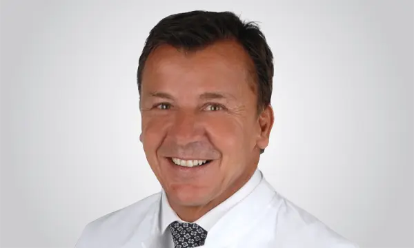
ATOS – Your spine specialist in Germany

At ATOS, you can expect cutting-edge medicine in many different areas including for spinal problems. Back pain is one of the most common ailments in the western world. Painful back problems are often due to problems with the spine. In terms of its functional role, on the one hand, the spine is responsible for keeping the body upright – it carries the weight of the head, trunk and arms – on the other, the spine protects the spinal cord.

Disc problems as a common back problem

The spine is divided into 7 cervical vertebrae, 12 thoracic vertebrae and 5 lumbar vertebrae. The sacrum and tailbone are also at the lower end. There are a total of 23 discs between the individual vertebrae which act as a kind of shock absorber. Wear, misalignment or accidents can cause the discs to shift and lose their elasticity. As a result, they can no longer act as shock absorbers between the individual vertebrae and the gelatinous nucleus of the intervertebral disc may shift (intervertebral disc bulging). If it breaks through the outer ring of the intervertebral disc it is called a herniated disc. If conservative therapies prove unsuccessful  intervertebral disc surgery is an option – in Germany, around 150,000 interventions are performed on intervertebral discs each year.

Back pain can have various causes

But intervertebral discs are not the only cause of pain in the back area. For example, a vertebral fracture is more common than commonly thought – and is not always noticed. In addition – especially in adolescents – spondylolisthesis is not uncommon. Overloading causes the vertebral bodies of the lower lumbar spine to move against each other – often accompanied by pain. If a doctor cannot control the back pain conservatively, surgery is inevitable. In old age, wear-related spinal canal stenosis also occurs – an increasing problem of our ageing society.

Our ATOS experts are among the most renowned spine specialists in Germany. Always on the cutting edge of research and technology, we can offer you spine treatments at the highest level.

You can rely on the expertise of our highly specialised and renowned physicians for back pain in our ATOS clinics.

Our treatments in detail

Intervertebral discs serve as a buffer between the vertebral bodies and function to cushion shocks. The interior is a soft and elastic gelatinous core, which is stabilised by a hard fibre core. If the fibre core gets cracked and the gelatinous core loses elasticity – both happens with advancing age – the danger of a herniated disc increases. This occurs when the gelatinous core slips due to excessive load and presses on the fibre core or breaks it.

Definition
By far the most common is a disc herniation (also called herniation or disc prolapse) occurring in the area of the lumbar spine (lumbar spine), because it carries a large load from our body. In far fewer cases, the cervical spine or thoracic spine is affected. Not only is age a contributing factor for a herniated disc of the cervical, thoracic, or lumbar spine, but obesity, predisposition, and improper loading, for example, by standing too long or sitting or by incorrectly lifting heavy loads are also factors. Disc herniation can therefore also occur in younger people.

Herniated disc at the thoracic spine
The thoracic spine in humans refers to the section between the cervical and lumbar spine. A herniated disc in the thoracic spine area is very rare. The causes of pain in the area of the thoracic spine should be examined in any case by a doctor because of the manifold possibilities. When diseases of the internal organs are excluded, then the causes are usually in the skeleton. Although disc herniations in the thoracic spine are far less common than lumbar disc herniations, in severe cases cross-sectional symptoms may develop and they may be associated with bladder or defecation problems. In addition to signs of wear and tear, nerve irritation due to bony injuries (fractures), blockages of the vertebral joints or inflammatory changes occur.

Symptoms – Herniated disc in the thoracic spine
Back pain in the area of the thoracic spine often occurs and is usually felt in a dull way between the shoulder blades or spreads in a belt shape over the chest. The affected area is usually sensitive to pressure.

Diagnosis – Herniated disc in the thoracic spine
An experienced doctor can already recognise postural defects with their own eyes. On the other hand, the use of imaging diagnostic procedures, for example magnetic resonance imaging (MRI), usually provides clarity about internal causes of pain. If there is a disc herniation of the thoracic spine, this is clearly visible in the tomograms. Often, however, so-called “thoracic spine pain” is also diagnosed as blockages of the inter-vertebral joints or rib-vertebral joints. In older people, a reduction in bone mass (osteoporosis) often causes ESPE pain, which can lead to vertebral body fractures with greatly reduced carrying capacity.

Conservative treatment – Herniated disc in the thoracic spine
The first therapeutic measure of a herniated disc is usually in the relief of acute pain by administering analgesics, which are usually also combined with anti-inflammatory or muscle relaxants. In most cases, improvements in discomfort can be achieved through physiotherapeutic therapies. Postural problems can be corrected by physical exercises and muscles can be specifically strengthened.

Surgical treatment – Herniated disc in the thoracic spine
Surgery for a thoracic disc herniation is more of an exception, and is usually only performed if the herniation of the thoracic spinal column presses on the nerves or spinal cord, or if there is a risk of paraplegia. Herniated discs at the level of the root to the side of the spinal cord are accompanied by excruciating pain. These disc herniations can be minimally invasively operated via a transforaminal approach.

Larger disc herniations, which involve a cross-sectional symptom, are operated via lateral access between the ribs. The vertebrae are then fused and bolted together from behind. Because it is a major surgery, surgery is only performed if paraplegia is imminent.

Rehabilitation – Time and methodology
Since 90% of herniated discs do not require surgery, the ultimate goal of rehabilitation is to eliminate pain and neurological discomfort. Rehabilitation is performed on an outpatient, semi-inpatient or inpatient basis. This depends on the severity of the symptoms. The drugs of choice in the treatments are:

  • Exercise therapy (stretching, strength, endurance)
  • Pain therapy with drugs or oral anesthesia (injection)
  • Psychological pain therapy for the decoupling of bad postures
  • Relaxation therapies
  • Back training for prevention
  • Occupational therapy
  • Nutritional advice to reduce body weight
  • Apparative processes (heat, electrical, ultrasound applications)

Normally, good results are achieved in 3-4 weeks.

If the related issues continue to be severe after 6-8 weeks (pain and dysfunction and, despite rehabilitation, there is no satisfactory improvement, surgery may be necessary. The rehab after an operation depends on the severity of the procedure. Experience shows that the patient should relax for the first 4-6 weeks after discharge. Only a moderate strain on the spine is recommended during this time. A rehabilitation programme with a specialist should only be started after this period.

Intervertebral discs serve as a buffer between the vertebral bodies and function to cushion shocks. The interior is a soft and elastic gelatinous core, which is stabilised by a hard fibre core. If the fibre core gets cracked and the gelatinous core loses elasticity – both happens with advancing age – the danger of a herniated disc increases. This occurs when the gelatinous core slips due to excessive load and presses on the fibre core or breaks it.

Definition
By far the most common is a disc herniation (also called herniation or disc prolapse) occurring in the area of the lumbar spine (lumbar spine), because it carries a large load from our body. In far fewer cases, the cervical spine is affected. Not only is age a contributing factor for a herniated disc of the cervical or lumbar spine, but obesity, predisposition, and improper loading, for example, by standing too long or sitting or by incorrectly lifting heavy loads are also factors. Disc herniation can therefore also occur in younger people.

The cervical spine forms the upper part of the human spine and consists of seven vertebrae. The neck also includes muscles, ligaments, bones and joints that are all crossed by nerves and respond to irritation or damage with severe pain. The intervertebral discs are located between the cervical vertebrae and act as a shock absorber for the body due to their elastic nature. They lose their elasticity with age or by stress, can tear when overstressed and (no longer) “soft” core can bulge out. The pressure on the spinal nerves or the spinal cord causes severe pain.

Symptoms – Herniated disc in the cervical spine
Neck and shoulder pain that may radiate into the arms, or numbness in the arms or hands, may be symptoms of a cervical disc herniation. But dizziness, headaches and tinnitus are reported by many sufferers. With chronic spinal cord injury (myelopathy), gait disturbances and other neurological dysfunctions also occur. They are in any case a sign that nerves in the neck or shoulder area are irritated by causes. These should be investigated by consulting a neurosurgeon.

Diagnosis – Herniated disc in the cervical spine
Our spine specialists examine your neck closely, pinpoint the pain centre and identify any movement restrictions. Of course, a thorough neurological examination is also included. X-ray examinations or magnetic resonance imaging (MRI) help our ATOS specialists diagnose the cervical spine disc herniation.

Conservative treatment – Herniated disc in the cervical spine
The pain and discomfort of the patient can be alleviated by medication and often also non-surgically by physiotherapeutic measures. If the herniated disc is in or at the root pocket and is associated with severe pain, a targeted radiological controlled injection to the nerve root (PRT) can be of great benefit. This can be offered as an outpatient service reliably in the clinic in cooperation with radiology.

Treatment surgically – Herniated disc in the cervical spine
The focus of surgical treatment of disc herniation in the cervical spine is the proven microsurgical ventral fusion using PEEK cage. A “cage” is a machine-shaped intervertebral space holder that replaces the damaged disc after it has been microsurgically removed. This cage restores the natural height of the intervertebral disc segment. PEEK is short for polyether ether ketone. It is a well-tolerated plastic in the body. The PEEK cage has a central cavity that fills with endogenous bone material after implantation.

An alternative to fusion of the vertebrae with cervical disc herniation is the insertion of a disc prosthesis, especially in younger patients with few signs of wear. An artificial disc preserves the mobility of the affected segment. It is constructed like a joint, the halves of which are each anchored in the adjacent vertebra. Most prostheses, depending on the manufacturer, consist of a plastic core and two outer metal plates that can be optimally anchored to the bone, so there is no risk of loosening.

Rehabilitation – Time and methodology
Since 90% of herniated discs do not require surgery, the ultimate goal of rehabilitation is to eliminate pain and neurological discomfort. Rehabilitation is performed on an outpatient, semi-inpatient or inpatient basis. This depends on the severity of the symptoms. The drugs of choice in the treatments are:

  • Exercise therapy (stretching, strength, endurance)
  • Pain therapy with drugs or oral anesthesia (injection)
  • Psychological pain therapy for the decoupling of bad postures
  • Relaxation therapies
  • Back training for prevention
  • Occupational therapy
  • Nutritional advice to reduce body weight
  • Apparative processes (heat, electrical, ultrasound applications)

Normally, good results are achieved in 3-4 weeks.

If the related issues continue to be severe after 6-8 weeks (pain and dysfunction and, despite rehabilitation, there is no satisfactory improvement, surgery may be necessary. The rehab after an operation depends on the severity of the procedure. Experience shows that the patient should relax for the first 4-6 weeks after discharge. Only a moderate strain on the spine is recommended during this time. A rehabilitation programme with a specialist should only be started after this period.
Intervertebral discs serve as a buffer between the vertebral bodies and function to cushion shocks. The interior is a soft and elastic gelatinous core, which is stabilised by a hard fibre core. If the fibre core gets cracked and the gelatinous core loses elasticity – both happens with advancing age – the danger of a herniated disc increases. This occurs when the gelatinous core slips due to excessive load and presses on the fibre core or breaks it.

Definition
By far the most common is a disc herniation (also called herniation or disc prolapse) occurring in the area of the lumbar spine (lumbar spine), because it carries a large load from our body. In far fewer cases, the cervical spine is affected. Not only is age a contributing factor for a herniated disc of the cervical or lumbar spine, but obesity, predisposition, and improper loading, for example, by standing too long or sitting or by incorrectly lifting heavy loads are also factors. Disc herniation can therefore also occur in younger people.

The causes of a herniated disc in the area of the lumbar spine are manifold and can also be genetically influenced. Often, the discs are damaged long before a herniated disc, and fluid loss and height reduction largely occur unnoticed. In many cases, abrupt twisting or flexing movements are the trigger for a fibre ring tear, which is exacerbated by risk factors such as work-related posture, as well as obesity, weak back muscles, or activities requiring a lot of sitting. Occasionally, a herniated disc occurs during pregnancy.

Symptoms – Herniated disc in the lumbar spine
Typically, there is severe pain in the affected area, which can radiate into the legs and usually disappears after some time. This pain occurs when an intervertebral disc is torn or damaged by overstressing. The deformed outer ring of the disc then presses on the spinal nerves, causing the pain. As all movement, coughing and sneezing intensify the pain, patients often experience cramped posture. The back muscles are reflexively hardened and blocked. Alarm signals include numbness or tingling, reduced reflexes, a sudden buckling of a leg, paralysis or unusual cold or heat sensations in the legs. The pain is often not precisely localised and affected patients indicated areas extending over 4-5 vertebrae. Most people complain of pain that extends into the buttocks, the leg or even into a foot, i.e. “lumbago”. Patients often cannot stand or walk on their heels or toes.

Diagnosis – Herniated disc in the lumbar spine
Our specialists first make a clinical report with special attention to the above signs of neurological impairment. For further proof, X-ray diagnostics, for example to exclude spinal gliding, is necessary. Subsequently, a magnetic resonance tomography (MRI) is indicated. This x-ray-free diagnosis makes it possible to detect a herniated disc with certainty. If necessary, additional neurological examinations may be required to assess the nerve conduction velocity of the affected segment.

Conservative treatment – Herniated disc in the lumbar spine
ATOS Clinics offer the entire spectrum of common conservative treatment measures for a lumbar disc herniation. This ranges from facet joint treatment using cryo -, heat or laser therapy of the vertebral joints to nerve root treatment under 3D X-ray. ATOS Clinics have state-of-the-art technical equipment for all of these treatments. In the acute phase, anti-inflammatory and often centrally effective pain medications are needed. Positioning measures (step bed), physiotherapy, manual therapy and local heat can relieve pain. Many patients benefit from these measures. The symptoms recede under treatment in eight to twelve weeks.

Surgical treatment – Herniated disc in the lumbar spine
If the muscles characteristically affected by the disc herniation can no longer be moved against gravity (strength degree 3 of 5 or less), there is an indication for surgery. Depending on the dynamics of the loss of strength of the muscle, it may even be an emergency situation that requires swift intervention. The same applies to the sudden occurrence of disorders in bladder and rectal control.

Herniated discs are operated on in a minimally invasive way using a microscope. Today, in contrast to earlier techniques, only the prolapsed material of the disc is removed. This is intended to preserve as much disc tissue as possible, which has an important shock absorbing function. The operation is performed prone with a small, approximately 3 cm long skin incision. Access to the spinal canal takes place between the vertebral arches while preserving the stability of the small vertebral joints. The spinal cord canal is then mobilised from the herniated disc and the prolapse recovered with micro-instruments. After surgery, the patient can immediately get up and walk around.

Rehabilitation – Time and methodology
Since 90% of herniated discs do not require surgery, the ultimate goal of rehabilitation is to eliminate pain and neurological discomfort. Rehabilitation is performed on an outpatient, semi-inpatient or inpatient basis. This depends on the severity of the symptoms. The drugs of choice in the treatments are:

  • Exercise therapy (stretching, strength, endurance)
  • Pain therapy with drugs or oral anesthesia (injection)
  • Psychological pain therapy for the decoupling of bad postures
  • Relaxation therapies
  • Back training for prevention
  • Occupational therapy
  • Nutritional advice to reduce body weight
  • Apparative procedures (heat, electrical, ultrasound applications)
    Normally, good results are achieved in 3-4 weeks.

If the related issues continue to be severe after 6-8 weeks (pain and dysfunction and, despite rehabilitation, there is no satisfactory improvement, surgery may be necessary. The rehab after an operation depends on the severity of the procedure. Experience shows that the patient should relax for the first 4-6 weeks after discharge. Only a moderate strain on the spine is recommended during this time. A rehabilitation programme with a specialist should only be started after this period.

The spinal cord and nerves pass through the spinal canal. Spinal stenosis is a narrowing of this canal, putting pressure on the nerves, spinal cord and blood vessels. This can result in pain and restricted mobility. Usually conservative measures are of little help with this disease. At ATOS Clinics, a competent medical team carries out the recommended operation as gently as possible.

Definition
The spinal canal is a kind of tunnel that runs from top to bottom through the spine. It consists of bone and connective tissue and protects the delicate spinal cord and nerve cords that run through the canal. If there is a constriction of the spinal canal, the nerves are constricted. The lumbar spine and the cervical spine are usually affected. Spinal canal stenosis is always progressive and cannot be stopped by the body itself. The reserve volume of the bony spinal canal required by the spinal cord and the spinal nerves is increasingly restricted and finally used up. The enlarged vertebral joints then protrude into the spinal canal and reduce the space for the nerve structures. Often, the bands that surround the spinal canal also thicken and also narrow it.

Symptoms
The symptoms associated with spinal canal stenosis in the lumbar spine usually begin with rather unspecific pain in the transition between the lumbar spine and the pelvis. Pulling in one or both buttocks halves or radiation of pain in the legs may occur. Standing for long periods feels unpleasant is avoided. Walking long distance is often also difficult. Unlike wearing in the hip (with pain onset) the first few metres are easy to handle. But after a short distance pain and sometimes numbness occur. There is also a feeling of tiredness. Patients often want to find a seat or walk in a bent position. This automatic “fix” indirectly expands the spinal canal and the nerve roots. This is caused by the small vertebral joints located at the back of the spine sliding apart. As a result, the patient’s movement is improved for a short time before the same symptoms recur. Patients usually have no problems riding a bicycle over longer distances. When shopping in the supermarket, they often use a shopping cart to hold themselves in a relaxed, bent-over position. Housework such as cooking or vacuuming, however, are poison for the patients because despite be bent over they are not supported.

Patients with spinal canal stenosis around the cervical spine increasingly lose their fine motor skills. Grasping and carrying objects is difficult. They report that objects fall out of their hands. Buttoning and unbuttoning clothing is difficult. They have trouble walking in the dark. Sudden turning movements are uncertain and require lunges to stabilise their upright posture. Clinically, in addition to the reported symptoms, both in the cervical and lumbar spine areas, weaknesses in the key muscles or later the loss of bladder and defecation control may occur.

Diagnosis
Diagnosis involves conventional X-ray diagnostics, which provides important static information about the alignment of the spinal column in space (balance), the development of wear-related scoliosis and the stability of the structure of the vertebral bodies (vertebral gliding). Furthermore, magnetic resonance imaging (MRI) is a central component of diagnostics. It provides high-resolution images of the bony and soft tissue narrowing of the spinal canal and can also reveal any negative changes in the spinal cord itself (myelopathy) at the cervical and thoracic vertebrae, which always lead to neurological deterioration in the short or medium term, if you do not do anything about it. If an MRI is not possible due to heart, brain pacemakers, or other metallic implants, a CT scan with contrasting of the spinal cord tubing may be performed to obtain the necessary information for treatment planning.

Conservative treatment
Therapy may be conservative in early stages without neurological deficits. Physiotherapeutic exercise therapies on a neurophysiological basis are used here to increase the coordination and fall prevention. Heat therapy can also have a favourable effect. Patients also benefit from manual lymphatic drainage.

Surgical treatment
The path to surgical therapy is often foreshadowed if the restrictions are too great or neurological impairments are impending. Patients should not wait to undergo surgical therapies until the deficit has occurred because it is never certain that it will regress. Surgical therapies should always be as minimally invasive as possible. Appropriate imaging is important for the planning. If sliding or significant decay of upright trunk posture (sagittal imbalance) can be ruled out, the bottlenecks revealed in the MRI can be microscopically supported and removed via small incisions without the use of implants. These techniques are also applicable to the cervical spine, as long as the tightness affects the root canal.

In case of symptomatic vertebral gliding or wear-related scoliosis, additional stabilisation in the same session may become necessary. This is done by us precisely with state-of-the-art, computer-aided navigation. As a result, the risk of implant failures can almost be ruled out entirely. The accesses can be minimised as well. We attach great importance to providing our patients with the greatest possible information on the operation, its preparation and the subsequent rehabilitation, as we are sure that the well-prepared patient will find their way back into their home and professional life faster.

The duration of the operation is about 1 hour.

Rehabilitation – Time and methodology
The patient can usually leave the clinic about 3 days after surgery. The rehabilitation phase consists of extensive physiotherapy treatment. A physiotherapist is visited and functional exercises are performed several times a week. The exercises help build muscles, which relieves burden on the spine. Furthermore, incorrect postures should be counteracted.

Vertebral gliding, known in the jargon as spondylolisthesis, is a sign of severe spinal instability. This can cause severe back pain and constriction of the spinal canal and threaten consequential damage. You should urgently consult a specialist if you experience neurological deficits. At ATOS Clinics we offer competent and experienced help with this medical issue.

Definition
Spondylolisthesis can be the result of natural degeneration of the spine. This means that as you get older due to prolonged stress and wear, the interplay of vertebrae, discs, joints, ligaments, and muscles gives way more and more. Loosening of the joints and ligaments, loss of elasticity of the intervertebral discs and weakening of the muscles result in slipping of vertebrae. Two or more vertebrae move relative to each other here. The displacement may occur rearward;  forward sliding, tilting and lateral displacement with rotation are also possible.

Spondylolisthesis occurs predominantly in the most stressed part of the spine, in the area of the 4th and 5th Lumbar vertebrae, but also occurs in other areas of the spine. The body tries to counteract the increasing instability caused by bony accumulation, which can lead to further complications such as narrowing of the spinal canal (spinal canal stenosis) and partly by a thickening of the joints (spondylarthrosis). In younger patients spondylolisthesis is mainly caused by a congenital bone weakness with defects in the area of the vertebral joints. A gap occurs between two vertebral bodies.

Symptoms
The sequelae and symptoms of degenerative spondylolisthesis include back pain that can radiate into the legs, movement restrictions in the spine, discomfort in the legs, loss of control of the bladder and bowel in case of nerve root stenosis or compression. Congenital spondylolisthesis, on the other hand, often causes little pain and is discovered incidentally during an x-ray examination. Spondylolisthesis can also cause a “caudal syndrome”. The syndrome must be recognised urgently; if untreated it could lead to permanent paraplegia.

Diagnosis
Vertebral gliding is readily visible in X-rays depending on the type and severity. In the sectional images of magnetic resonance imaging (MRI), the pain occurring can be additionally assigned to organic areas. Likewise, damage to soft tissues such as intervertebral discs, nerve roots and ligamentous structures can be visualised in an MRI. Our spine specialists at ATOS Clinics provide you with all the prerequisites for a reliable diagnosis through many years of experience and state-of-the-art technical equipment.

Conservative treatment
One can initiate the therapy conservatively with the following approaches. This is called a conservative modular therapy concept:

  • Physiotherapy (movement therapy with baths and muscle-relaxing treatments) to relieve and stabilise the spine
  • Electrotherapy for pain treatment and muscle relaxation
  • Support corsets (orthoses) to relieve the spine
  • Back training with targeted strength training for the back and abdominal muscles
  • Targeted pain therapy

Surgical treatment
In order to fix the gliding permanently, however, an operation is usually necessary. In vertebral displacements with compression of nerves and instability, fusion of the affected vertebrae (spondylodesis) is a possible solution. We prefer percutaneous fusion at ATOS Clinics.

In a percutaneous fusion, the vertebral bodies are adjusted and fixed with minimally invasive techniques under X-ray monitoring by means of screws and rods. With this surgical technique, the size of the skin incisions and the scarring on the large back muscles are significantly reduced compared to conventional open surgeries. Less traumatisation of the soft tissue close to the spine leads to less postoperative pain, which results in a short hospital stay and faster rehabilitation.

The percutaneous fusion of the spine has proven its value in cases of primary or secondary instability, when no progress can be achieved by conservative treatment. By stabilising the spine, the back pain caused by the damaged disc in case of instability is suppressed.

In most cases, minimally invasive techniques are used. The duration of the operation is about 1 hour.

Rehabilitation – Time and methodology
The rehab can usually begin 8-12 weeks after surgery. The body needs rest after surgery until the new foreign bodies in the spine are ossified. Subsequently, the stabilising corset is gradually no longer used and physiotherapeutic exercises for the strengthening of the back muscles is started.

A vertebral fracture is a fracture of a vertebral body of the spine. The vertebral fracture can occur in many different forms. Vertebral fractures account for up to two percent of all fractures and can have serious effects. At ATOS Clinics our spine specialists are at your disposal with modern, technical medical equipment for confident vertebral fracture diagnosis. We can even treat complicated fractures.

Definition
The spine provides the right support in the human body and protects internal organs as well as the spinal cord. It consists of different parts, which include the vertebral bodies. They are actually a stable bone fragment. Nevertheless, they can also break, for example, if they are struck. The trigger for a vertebral fracture may be a large external force or a fall with a simultaneous rotation of the spine, for example in a motorcycle accident or a fall from a high altitude.

If the vertebral bones are damaged by osteoporosis and are unstable, even lower forces are sufficient to break the vertebrae. A vertebral fracture is dangerous in any case because the spine loses its stability. There is a risk that parts of the brittle vertebra may shift and even injure the spinal cord. If a vertebral fracture is suspected, a specialist should be consulted for a clear diagnosis.

Symptoms
A vertebral fracture can lead to very different complaints. They depend on the cause of the injury. An osteoporotic vertebral fracture causes, for example, permanent back pain that cannot be clearly defined. In contrast, vertebral fractures caused by trauma cause immediate pain at the corresponding site.

Frequently affected people only go to the doctor when pain occurs. When the fracture presses on the surrounding structures, various symptoms may occur. They can range from paralysis in the legs to impaired sensation of touch.

Diagnosis
In the event of pain or after a serious accident vertebral injury is suspected, an experienced specialist should get to the bottom of the matter. This ensures consequential damages can be excluded. A vertebral fracture can clearly diagnosed with magnetic resonance imaging (MRI). At ATOS Clinics, we discuss the exact diagnosis in detail with the patient, clarify all their questions and together decide on the further steps for optimal treatment.

Conservative treatment
Many physicians recommend conservative therapies with immobilisation and wearing a corset in the event of a slight vertebral fracture due to osteoporosis. Our ATOS specialists instead recommend a vertebral fracture operation. The advantages are that the patient can be relieved of the acute pain and the risk of spinal cord pinching can be better addressed. It should be noted that surgical spinal procedures have evolved considerably and, in most cases, are no more risky than other surgeries.

Surgical treatment
At ATOS Clinics, an experienced team of specialists is ready to treat even complicated fractures. Of course, we also support you during vertebral rehab and jointly develop the perfect programme for your speedy recovery.
There are currently three types of operations:

  • Percutaneous balloon kyphoplasty
    In percutaneous balloon kyphoplasty, a medical balloon is inserted through a small incision in the vertebral body on both sides. It is then inflated with contrast agent to create a cavity. Subsequently, the cavity is filled with bone cement, whereby the vertebra is stabilised after curing. This very gentle vertebral fracture surgery method has almost completely displaced the outdated vertebral plasty (injection of cement without forming a cavity in advance and without stabilising the fractured vertebra).
  • Percutaneous spondylodesis
    In the case of percutaneous spondylodesis (fusion), the vertebral bodies above and below the fracture are adjusted and fixed with a minimal invasive technique under X-ray monitoring by means of screws and rods. With this surgical technique, the size of the skin incisions and the scarring on the large back muscles are significantly reduced compared to conventional open surgeries. Less traumatisation of the soft tissues close to the spine results in less postoperative pain, which usually allows for shorter hospital stays and faster rehabilitation.
  • Combination of balloon kyphoplasty and spondylodesis
    With instabilities, fusion of two or more vertebral bodies is often necessary in combination with the aforementioned methods. In all cases described above, our doctor will discuss the optimal vertebral fracture therapy for your individual diagnosis in detail with you, give you comprehensive advice and answer all questions about the procedure, the benefits and the risks as well as side effects.

Rehabilitation – Time and methodology
The healing time of a vertebral fracture depends heavily on how severe the trauma is. A stable vertebral fracture usually solidifies after successful surgical intervention in a few weeks. It becomes solid and stable again. Patients may mobilise (depending on the level of pain) immediately or after about 3 weeks. In case of unstable fractures and cervical vertebra fractures, healing can take between 6-9 months.

Your spine specialists at ATOS Clinics

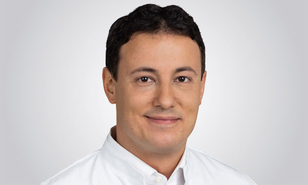
All our doctors have many years of experience. Find your specialist here and make an appointment. The order of our doctors in the following is purely alphabetical and does not represent a qualitative ranking.