ATOS – Your hand specialist in Germany

Our ATOS experts offer top medical care in every respect – for hand problems as well. The human gripping tool consists of 27 bones. It is formed from the metatarsal root, fingers and a variety of muscles, tendons and nerves. The hand is movable and can be used very precisely due to this variety of different components. But its delicate structure also makes the hand susceptible to wear and injuries.

Hand orthopaedics is specialised care

Hand orthopaedics includes the treatment of diseases, injuries or malformations of the fingers, hands, wrists and forearms up to the elbow (tennis elbow). Whether it is minor hand injuries, rhizarthrosis, or complex nerve dysfunction syndrome (carpal syndrome): The medical team at ATOS Clinics, our specially trained nursing staff, our innovative conservative and operative therapies and last but not least our medical equipment make our clinics a leading European expertise centre for hand orthopaedics.

Our ATOS experts are among the most renowned hand specialists in Germany. Always on the cutting edge of research and technology, we can offer you hand treatments at the highest level.

You can rely on the expertise of our highly specialised and renowned physicians for hand pain in our ATOS clinics.

Our treatments in detail

Dupuytren’s disease is a connective tissue disease of the hand and fingers, causing increasing flexion of the fingers. Extending the fingers becomes more difficult and ultimately impossible.

Definition
Dupuytren’s disease was named after it discoverer, the French physician Baron Guillaume Dupuytren, and is usually noticeable through the formation of tight knots in the cupped hand. Dupuytren occurs mostly in middle age and in men earlier than in women. Typically, the small and ring fingers are affected, more often on the right hand than on the left hand. The disease affects about 1.5 million people in Germany alone. A definite cause is unknown. Familial accumulation is observed, but also metabolic diseases or intoxications are suspected, but so far have not been proven as causes. In the long term, only a surgical intervention by a hand specialist can help with this disease. At ATOS Clinics, the pathologically altered tissue and thus the cause of the disease is surgically and painlessly removed.

Symptoms
A tissue change (fibrosis), which causes a pathological increase of connective tissue cells is responsible for the hard knots. Over time, the knots develop into hardened strands in the fingers, which initially bend the base joints of the fingers and later also the middle joints. Extending the fingers becomes more difficult and ultimately impossible. The disease develops relatively slowly, usually painless and is therefore hardly noticed at first.

Diagnosis
The doctor scans the palm of the hand to sense nodular changes or hardening on the palm or along the sides of the fingers. He then moves your fingers one at a time to assess the strength of the movement restrictions. To rule out other conditions, an X-ray or magnetic resonance image may be necessary.

Conservative treatment
Dupuytren is often treated with conservative therapies such as laser radiation, gymnastic exercises, shock-wave therapy, etc. However, the effectiveness of these methods is controversial. For this reason, our hand specialists at ATOS Clinics recommend a Dupuytren’s operation to permanently eliminate the condition.

Surgical treatment
Delays due to conservative treatment of the disease can result in complete collapse of the fingers with low supply and infection. Only the operative removal of the contracting strands and the diseased fascia tissue has proven successful. The Dupuytren’s operation is usually performed only when the disease has caused movement restrictions.

The duration of the operation and whether it is performed on an outpatient or inpatient basis depends on the severity. This surgical procedure often produces a large wound on the inside of the hand. For this reason, we consider it necessary to perform wound monitoring and care at short intervals in the first one to two days after surgery, so we recommend our patients stay in hospital for one to two days.

Rehabilitation – Time and methods
After the surgical procedure, wound healing takes place over a period of 3-6 weeks. Intensive follow-up with 2-3 changes of bandage weekly in the first two weeks and removal of the dermal stitches after about 10 days is recommended.

Osteoarthritis refers to the wear of the articular cartilage. In the area of the wrist, this usually represents the long-term consequence of an inflammatory joint disease (rheumatoid arthritis) or an accident. Poorly healed fractures (distal radius fracture, scaphoid fracture) or ligament injuries (ligament injury) can lead to disturbed biomechanics or instability. These accelerate the joint wear and promote osteoarthritis in the hands, which is accompanied later by wrist pain.

Definition
The wrist is structured differently than the shoulder and hip. The joint does not consist of a head and socket. It is actually a joint area with cartilage between the radius and carpus. That means cartilage wear or complete deformation is possible in every area of the wrist. In most cases, wrist arthrosis is located between the radius and the carpal bones.

Symptoms
In the early stages of arthritis in the hand, patients report load-related symptoms and a tendency to swell. Later on there is usually a restriction of movement and also increasing resting pain and nocturnal pain in the wrist. “Morning pain” with joint stiffness that decreases during the day is also characteristic of wrist arthrosis.

Diagnosis
The diagnosis of arthritis in the hand can be confirmed by a simple x-ray of the wrist.

Conservative treatment
There is no drug therapy for the restoration of cartilage; therefore, the main goals of a treatment are pain relief and mobility preservation. The focus of a conservative treatment is:

  • Manual therapy
  • Physiotherapeutic exercises
  • Heat applications such as ultrasound and hand baths

Surgical treatment
Surgical procedures are always used in wrist arthrosis when conservative treatment methods can no longer provide adequate pain relief. Depending on the situation, ligament injuries (SL band) can lead to instability in the wrist area, which can lead to cartilage wear and arthrosis between individual carpal bones. It is possible in many cases here to remove only the affected carpal bones and perform a partial fusion. So residual mobility can be retained.

In mediocarpal partial arthrodesis, the navicular bone is removed and a partial fusion of four carpal bones is performed. The articular surface between the lunate bone and radius is preserved and guarantees good function with significant pain relief. Alternatively, removal of the proximal carpal row (PRC) may also be considered here.

In the case of advanced arthritis with destruction of the articular surfaces, a painful restriction of movement (wobble stiffness) has already developed. The surgical fusion fixes the painful residual mobility and achieves a stable, low-pain situation. During surgery, the destroyed articular surfaces between the distal radius and the carpus are dissected and the remaining cartilage is removed. A stable bone block is usually made with a stable titanium plate. Due to the fusion, few disadvantages occur in everyday life, and the turning movement of the forearm is not significantly limited after the operation.

Rehabilitation – Time and methods
The prognosis for a healing process depends on the bone quality. Should a dense bone block form directly it can be assumed that there will be a great deal of freedom from complaints. Smokers have a statistically greater risk of failure of bones to heal. After the operation, the wrist is splinted on the outside. It takes several weeks for the bone to heal completely.

Carpal tunnel syndrome is one of the most common causes of hand pain and discomfort. This nerve palsy disorder of the median nerve occurs in the wrist and manifests itself in pain and motor disorders in the hand. Often the hand can no longer be clenched into a fist, and fine motor skills suffer.

Definition
The arm’s three major nerve cords have to pass through several anatomical bottlenecks on their way from the cervical spine into the fingers. In the passages through bony channels or muscles and tendons, the nerves can be irritated for various reasons and cause pain. Sensations such as tingling fingers or numbness of the fingers or the hand and weakness are some symptoms of nerve constriction syndromes. The most common nerve dysfunction syndromes are Carpal Tunnel Syndrome (KTS) and Ulnar Disease Syndrome (Sulcus Ulnaris).

One of the anatomical bottlenecks in the arm is the so-called carpal tunnel in the wrist. In the carpal tunnel, nine tendons pass from the forearm into the hand and to the median nerve. Carpal bones limit this tunnel on the sides and bottom. At the top, the carpal strap closes the rather narrow carpal tunnel. If there is an increase in volume in the tunnel, the sensitive central nerve is pressed. And if the pressure persists it causes painful irritation. The reasons for the increase in volume may be swelling of the hand or forearm, inflammation of the tendons, broken bones in the hand or tendinitis.

Symptoms
The most typical sign of carpal tunnel syndrome is (at an early stage) the fingers falling asleep in the evenings. Very severe pain occurs later. The pain can then spread to the arm and shoulder. In the morning, the fingers are often swollen and stiff.

Diagnosis
Our specialist for hand and arm discomfort examines the hand using various tests in order to diagnose carpal tunnel syndrome after the patient interview (anamnesis) and takes X-rays if necessary. Damage can also be detected by measurements of nerve conduction velocity, whereby the nerve constriction syndrome can also be located and assessed for severity.

Conservative treatment
With a mild form of carpal tunnel syndrome, the discomfort can be alleviated by medication and by immobilising the hand with a wrist splint. Other possible treatment options include ultrasound, kinesio tape and acupuncture.

Surgical treatment
If the symptoms are too severe, an operation is necessary in which the splitting of the tendon structure around the carpal tunnel reduces the pressure on it. Such surgery can usually be done arthroscopically. Minimally invasive carpal tunnel syndrome surgery makes it easier for older people to walk again with walking sticks or walking aids.

Rehabilitation – Time and methods
After the carpal tunnel operation the hand does not need to have a plaster cast placed on it. That means the fingers can be used right away. The operated hand should be rested for at least 14 days. The duration also depends on the extent of pre-existing nerve damage. The connective tissue of the carpel roof heals postoperatively in 6-10 weeks. Afterwards the patient can place a full load on the hand and resume sports activities.

Pain in the thumb often indicates thumb saddle joint osteoarthritis – medically referred to as rhizarthrosis. It causes pain and hinders gripping and holding movements of the thumb and thus the hand. Our specialists in hand orthopaedics at ATOS Clinics offer a whole package of proven and successful therapies for rhizarthrosis.

Definition
The term rhizarthrosis comes from the Greek “rhiza”, which means “root”. It refers to arthritis of the thumb saddle joint and is one of the most common signs of wear of the joints of the hand. Abrasion of the protective cartilage layer in the joint creates additional friction that causes painful inflammation and swelling. Ultimately bone rubs on bone, and the joint is destroyed.

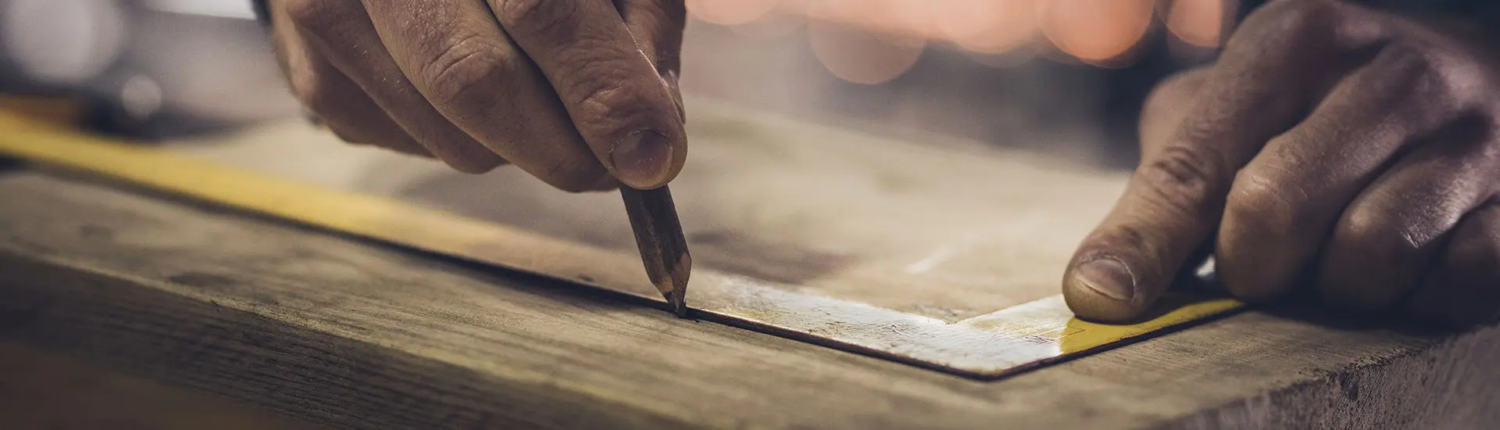
The thumb saddle joint is one of the most stressed joints in humans and is greatly strained by the large number of sometimes very strong gripping and holding movements. For artisans such as tilers or plumbers, rhizarthrosis is considered an occupational disease. It is a hereditary condition, occurring in women more often than in men.

Symptoms
The severity of the pain depends on the stage of osteoarthritis and varies within an astonishing range. There are patients who do not complain of pain even with severe arthrosis, but also patients who already experience severe pain with mild osteoarthritis. In addition, pain often occurs only after heavy strain on the thumb.

Diagnosis
Diagnosis of thumb saddle joint arthrosis begins with a medical history report and medical examination. It is confirmed by x-rays. An examination for a possible diagnosis must be carried out with additional feelings of numbness.

Conservative treatment
There is no drug therapy for the restoration of cartilage; therefore, the main goals of a treatment are pain relief and mobility preservation. With conservative therapies such as a thumb splint, ice treatment, medication or electrotherapy, the symptoms can be alleviated. This can also be achieved through acupuncture, injections of hyaluronic acid and mixed corticoid injections.

Surgical treatment
In the event of persistent symptoms, the pain and restrictions of movement can be permanently removed by various surgical therapies:

Denervation
With a denervation, the pain-transmitting nerve fibres are interrupted. However, this operation only works for a few years as the nerve fibres regrow.

Resection arthroplasty
With so-called resection arthroplasty, the arthritic parts of the carpal bone are removed and replaced by tendon surgery for improved stability.

Fusion operation (arthrodesis)
In younger patients or artisans, a fusion operation (arthrodesis) is often preferred, which offers the advantage of strong resilience of the joint at the expense of fusing the thumb joint.

Rehabilitation – Time and methods
After the thumb joint osteoarthritis surgery, the thumb is immobilised for about 2 weeks. In some cases, the immobilisation must be extended to 4-6 weeks. After removal the sutures and with good wound healing careful load increase can be started under the guidance of a physiotherapist.

ATOS – Your specialist for hand diseases in Germany